
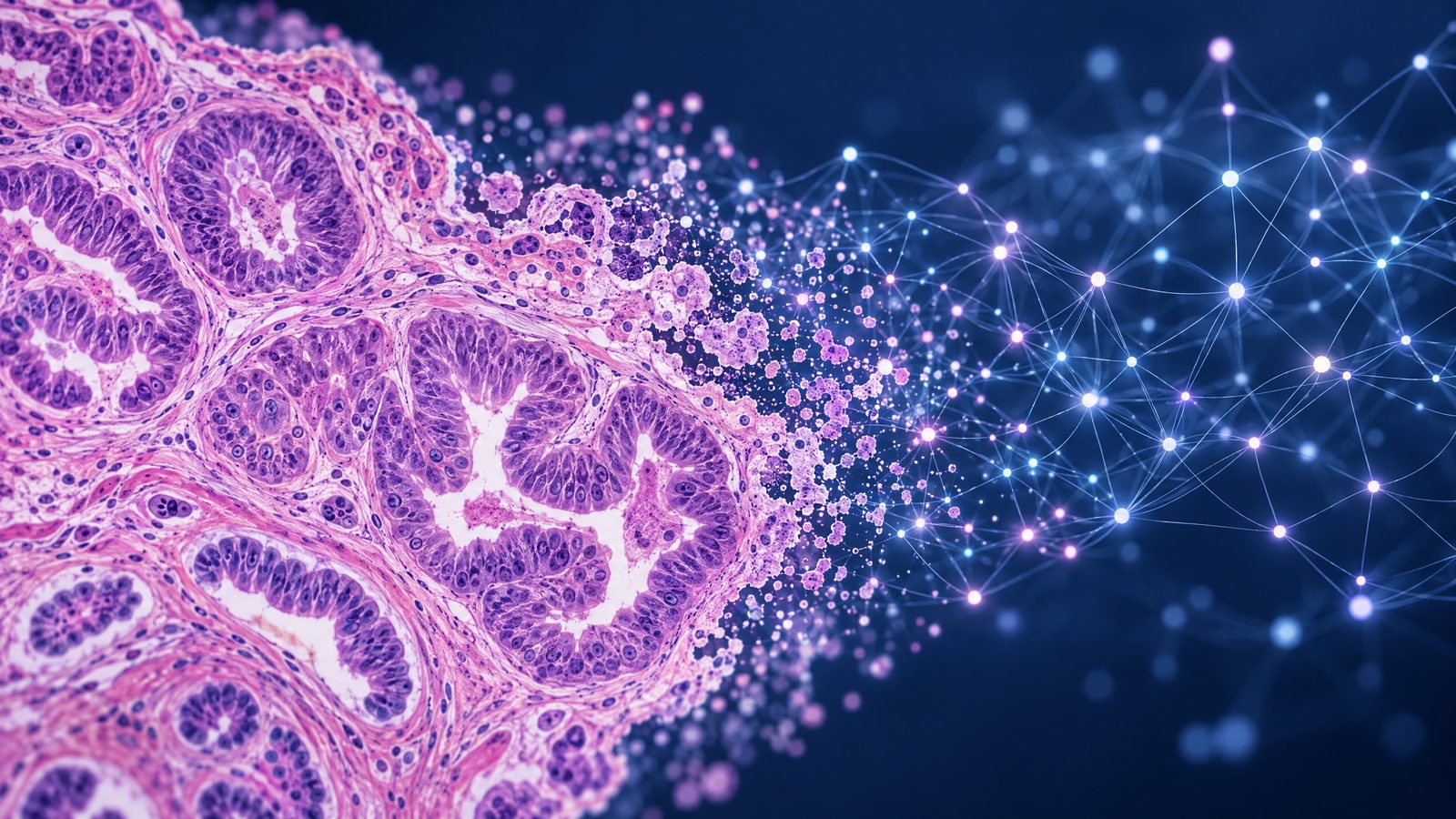

The research
Researchers at King’s College London have reported an experimental artificial-intelligence model that looks for signs of faulty DNA repair in ovarian cancer tissue images. The model, called HRDPath, was designed to predict homologous recombination deficiency, or HRD, in high-grade serous tubo-ovarian carcinoma. This cancer can arise in the ovaries or fallopian tubes. The study was published on 21 October 2026.
HRD matters because it can help inform treatment choices. Tumours with this characteristic have impaired ability to repair certain DNA damage and may be more vulnerable to platinum-based chemotherapy and PARP inhibitors. At present, assessment relies on genomic testing. The research explores whether routinely prepared microscope images might provide another source of clues; it does not establish that image analysis can replace laboratory testing or guide care on its own.
How the model was assessed
HRDPath analyses digitised whole-slide images of tissue. The researchers trained it using samples for which HRD status had already been assessed through genomic analysis, then tested performance across separate patient groups. The work included an in-house cohort and two public datasets, with validation extending beyond the material used to develop the model.
The institutional summary reports that when several images from a patient were considered together, the model correctly identified approximately 91 per cent of HRD-positive cases. That figure describes the share of positive cases detected in that setting; it is not an overall accuracy rate, nor does it mean that nine in ten patients would receive a correct clinical result. The paper also reports performance under other evaluation conditions, underscoring that results depend on how images and cases are assembled. A single headline percentage cannot convey false positives, false negatives or how performance might vary in another hospital population.
What the images may reveal
The researchers examined image features associated with HRD as well as the model’s predictions. They describe differences in cell appearance and tissue organisation, including rounder cell nuclei and more densely packed cancer cells in some tumour regions. These are associations observed in the study, not a basis for diagnosing an individual from appearance alone.
A potential attraction of the approach is that pathology images are already produced during routine cancer assessment. If future studies establish that image-based predictions are reliable across settings, the technique might help identify cases for further testing or research and could eventually widen access to biomarker information. Those possibilities remain prospective. The study did not demonstrate that patients can safely skip genomic tests, that treatment decisions were improved, or that the model is available for clinical use.
Validation remains essential
The findings are an early research result, not a validated diagnostic service. The researchers say the model needs validation in larger patient populations. That work would need to establish how reliably it performs across hospitals, scanners, tissue preparation methods and different patient groups, and how often it misses HRD-positive cancers or flags HRD where it is absent. The performance of any future system would also need to be assessed against the existing genomic methods used to establish HRD status.
The study therefore points to a possible way of extracting additional information from tissue already examined under a microscope, rather than proving a shortcut to treatment selection. For now, HRDPath should be understood as an investigational model. Its reported ability to identify many HRD-positive cases when multiple images are combined is a reason for further evaluation, not evidence that it is ready to make clinical decisions.